Human Skin Cell Atlas: What Real Cell Data Reveals About Skin Repair
Can a scientific map of human skin help researchers understand wound healing, hair follicles and future treatments? The Human Skin Cell Atlas is based on real measurements, but its scientific purpose is more precise than
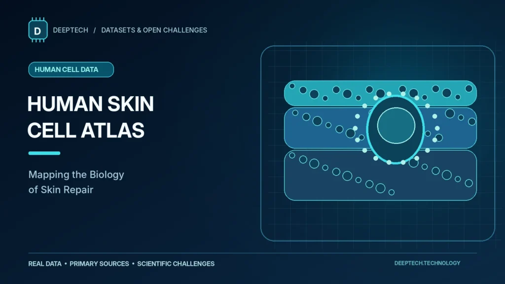
Original conceptual editorial illustration; not a research microscopy image.
Can a scientific map of human skin help researchers understand wound healing, hair follicles and future treatments? The Human Skin Cell Atlas is based on real measurements, but its scientific purpose is more precise than a promise to regrow or rejuvenate skin.
What this dataset can actually verify
An atlas built from many single-cell studies has two distinct forms of evidence: the pooled cell profiles and a smaller number of tissue sections with spatial measurements. The GEO record GSE308929 describes the integrated 34 source datasets and separately documents two spatial sections from healthy facial skin. Those two sections came from the same donor. They offer useful anatomical information, but do not count as independent validation across different people.
This matters when comparing the atlas to a clinical claim. A cell cluster reproducibly observed across independently collected studies would be stronger evidence of a broadly shared cell state than a marker found in only one laboratory batch. Spatial measurements can help check where a cell state appears, but one donor cannot establish how consistently that arrangement varies with age, sex, skin condition or body site.
Why this scientific question matters
Skin is a protective organ that continuously renews itself. Chronic wounds, inflammatory diseases and scarring can disrupt that process. To understand possible therapeutic strategies, researchers first need reliable knowledge of which cells exist in healthy tissue, where they are located and how they communicate. A cell atlas is a reference map for asking those questions rather than an intervention administered to a patient.
The pilosebaceous unit, which includes hair follicles and associated glands, is particularly complex. It participates in skin maintenance and disease, yet some of its deeper structures have been less comprehensively described than more accessible skin layers. Mapping these structures accurately may eventually help researchers investigate why some tissue responses become pathological.
What the researchers actually measured
The research article An integrated skin cell atlas decodes the pilosebaceous unit, made available in the Journal of Investigative Dermatology in July 2026, describes integration of 34 previously published single-cell RNA-sequencing datasets comprising 818,951 cells. Its public GEO repository summary reports 819,068 cells. DeepTech preserves both figures because the source records differ; a reanalysis should first check the respective filtering and reporting definitions.
The project also conducted high-resolution spatial transcriptomics on healthy adult human facial skin samples, locating gene-expression patterns within tissue sections. The GEO entry documents the samples and data formats. These are laboratory observations, not artificially generated clinical cases.
How could this inform treatments?
Healthy reference atlases may help scientists identify cell markers, signalling relationships and candidate pathways worth studying in wound repair or skin disorders. They can also provide a baseline against which future diseased or injured samples can be compared. These are plausible research applications grounded in the authors’ emphasis on tissue organisation, homeostasis and pathology.
However, a pattern in gene-expression data does not establish that activating or suppressing it will produce safe skin repair. A proposed therapy would need functional cell or tissue experiments, appropriate animal or other preclinical testing, and, if applicable, clinical studies.
Five research challenges worth attempting
- Can independently collected skin samples reproduce the same cell-type annotations?
- Which markers are stable across age, body sites and laboratories?
- Can spatial measurements verify relationships inferred from single-cell profiles?
- Do disease-associated signals differ consistently from healthy reference tissue?
- Can the source data and each analysis decision be traced and reproduced?
Where to inspect the source data
NCBI GEO accession GSE308929 contains the public deposit, its samples and links to downloadable data. The peer-reviewed study provides the research context and reported findings. Researchers should review reuse terms, donor metadata and methods before drawing comparisons.
Explore more primary-source projects in the DeepTech Datasets & Open Challenges directory. This article is scientific reporting; it does not claim that a cure or regenerative treatment has been demonstrated.
A concrete reproducibility protocol
A researcher could begin by downloading the processed cell-by-gene matrix and harmonised labels, recording the accession, file versions and filtering decisions. The first analysis would reproduce a published marker map without changing the cell types after seeing the result. Next, one contributing dataset could be withheld completely during atlas construction and used only to assess whether the marker set recurs in independently collected samples. The withheld dataset must not influence which markers are chosen.
- Unit of independence: compare donor and study identifiers; thousands of cells from one donor are not thousands of biological replicates
- Distribution shift: report which body sites and sample-preparation methods are represented in the training and evaluation material
- Spatial follow-up: inspect the two deposited skin sections, but do not treat them as a separate multi-donor validation cohort
- Interpretation: describe a molecular association as a hypothesis until a functional experiment tests the proposed biological effect
DeepTech has not executed this protocol. Its purpose is to show readers exactly which independent checks would strengthen the reported atlas, while respecting donor privacy and the original study’s limits.